The Cell Cycle and Radiosensitivity: Essential Concepts for FRCR Part 1
Master the cell cycle phases, checkpoint regulation, and cell cycle-dependent radiosensitivity for FRCR Part 1. Understand how radiotherapy exploits cell cycle biology.
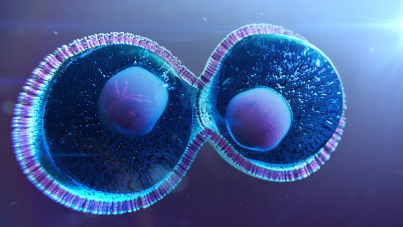
Understanding the cell cycle is fundamental to radiobiology and oncology. Cell cycle phase determines radiosensitivity, checkpoint function affects DNA damage response, and checkpoint dysregulation is a hallmark of cancer. This guide covers the essential concepts for FRCR Part 1.
Cell Cycle Phases Overview
The cell cycle consists of four main phases:
G1 (Gap 1) Phase:
- Cell growth and protein synthesis
- Preparation for DNA replication
- Decision point: proceed to division or exit to G0
- Duration: variable (hours to days)
S (Synthesis) Phase:
- DNA replication occurs
- Chromosome number doubles (2N to 4N DNA content)
- Duration: typically 6-8 hours
G2 (Gap 2) Phase:
- Preparation for mitosis
- Continued protein synthesis
- Final checkpoint before division
- Duration: typically 2-4 hours
M (Mitosis) Phase:
- Nuclear division (karyokinesis): prophase, metaphase, anaphase, telophase
- Cell division (cytokinesis)
- Duration: approximately 1 hour
G0 Phase:
- Quiescent state (cells exit the cycle)
- Terminally differentiated cells remain here permanently
- Some cells can re-enter the cycle if stimulated
Cell Cycle Regulation: Cyclins and CDKs
Cell cycle progression is driven by cyclin-dependent kinases (CDKs) which require binding to cyclins for activation:
Key Cyclin-CDK Complexes:
- Cyclin D - CDK4/6: G1 progression, phosphorylates Rb protein
- Cyclin E - CDK2: G1/S transition
- Cyclin A - CDK2: S phase progression
- Cyclin B - CDK1 (CDC2): G2/M transition, mitosis entry
The cyclin-CDK complexes phosphorylate target proteins to drive the cycle forward.
Cell Cycle Checkpoints
Checkpoints are surveillance mechanisms that ensure proper cell cycle progression:
G1/S Checkpoint (Restriction Point):
- Major decision point for cell cycle commitment
- Checks for DNA damage, adequate cell size, growth signals
- Regulated by Rb and p53
- Most frequently dysregulated in cancer
Intra-S Phase Checkpoint:
- Monitors ongoing DNA replication
- Responds to replication stress or damage
- ATR kinase is the primary sensor
G2/M Checkpoint:
- Ensures DNA replication is complete before mitosis
- Checks for DNA damage
- ATM/ATR → Chk1/Chk2 → Cdc25 inhibition → CDK1 inactive
Spindle Assembly Checkpoint (Metaphase):
- Ensures all chromosomes are properly attached to spindle
- Prevents aneuploidy
DNA Damage Checkpoint Signalling
When DNA damage occurs, a signalling cascade is activated:
- Sensors detect damage (MRN complex, RPA)
- Transducer kinases are activated (ATM for DSBs, ATR for replication stress)
- Effector kinases propagate signal (Chk1, Chk2)
- Cell cycle arrest occurs through Cdc25 inactivation and p53 stabilisation
p53 - "Guardian of the Genome":
- Normally kept at low levels by MDM2-mediated degradation
- DNA damage causes p53 phosphorylation and stabilisation
- p53 induces p21 (CDK inhibitor), causing G1 arrest
- Can also trigger apoptosis if damage is irreparable
- p53 is mutated in >50% of human cancers
Cell Cycle and Radiosensitivity
Cells show variable radiosensitivity depending on cell cycle phase:
Most Radiosensitive:
- M phase (mitosis): Chromosomes are condensed, no repair occurs
- G2 phase: Limited time for repair before mitosis
Intermediate Sensitivity:
- G1 phase: Time available for repair
Most Radioresistant:
- Late S phase: Homologous recombination repair available (sister chromatid present)
- Early S phase: Also relatively resistant
Clinical Implications:
- Fractionation allows redistribution through the cell cycle (one of the 5 Rs)
- Cells in resistant phases during one fraction may be in sensitive phases during subsequent fractions
Cell Cycle Dysregulation in Cancer
Cancer cells typically show:
- Loss of G1/S checkpoint (p53/Rb dysfunction in >90% of cancers)
- Increased reliance on G2/M checkpoint for DNA damage response
- This creates therapeutic vulnerability to G2/M checkpoint inhibitors
Common Alterations:
- p53 mutations (~50% of cancers)
- Rb pathway dysfunction (CDK4/6 amplification, cyclin D overexpression, p16 loss)
- Cyclin E amplification
Therapeutic Targeting
CDK4/6 Inhibitors:
- Palbociclib, ribociclib, abemaciclib
- Block G1 progression in Rb-intact cells
- Used in hormone receptor-positive breast cancer
Checkpoint Kinase Inhibitors:
- Chk1/Chk2 inhibitors under investigation
- Wee1 inhibitors (adavosertib)
- May sensitise p53-deficient tumours to chemotherapy/radiotherapy
Key Exam Points
- Cell cycle phases: G1 → S → G2 → M (G0 = quiescent)
- Cyclin D-CDK4/6: G1; Cyclin E-CDK2: G1/S; Cyclin B-CDK1: G2/M
- Most radiosensitive: M and G2 phases
- Most radioresistant: Late S phase
- p53 induces G1 arrest via p21 (CDK inhibitor)
- ATM senses DSBs; ATR senses replication stress
- G1/S checkpoint most commonly dysregulated in cancer
- Redistribution through cell cycle is one of the 5 Rs of radiobiology
Enjoyed this article?
Share it with a colleague preparing for their FRCR.