The TNM Cancer Staging System: Essential Principles for FRCR Part 1 Success
Understand the principles of TNM staging, AJCC 8th edition updates, and clinical applications for FRCR Part 1. Learn how anatomic staging integrates with molecular factors in modern oncology.
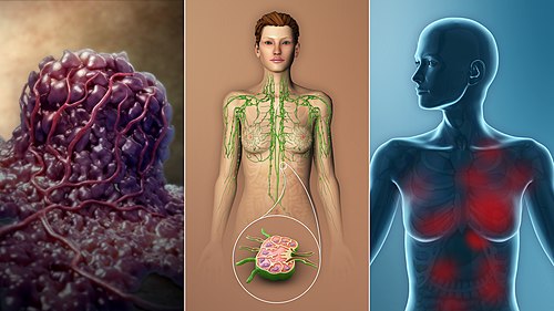
The TNM classification system is the international standard for cancer staging, defining prognosis and guiding treatment decisions. For FRCR Part 1 candidates, understanding staging principles is essential, as staging concepts appear across all oncology modules.
History and Development
The TNM system was first developed by Pierre Denoix in the 1940s and 1950s. The American Joint Committee on Cancer (AJCC) published its first staging manual in 1977, with the 8th edition released in October 2016 (implemented January 2018). The Union for International Cancer Control (UICC) maintains compatibility through collaborative development.
Fundamental TNM Components
T - Primary Tumour:
- TX: Primary tumour cannot be assessed
- T0: No evidence of primary tumour
- Tis: Carcinoma in situ
- T1-T4: Increasing size and/or local extent
N - Regional Lymph Nodes:
- NX: Regional nodes cannot be assessed
- N0: No regional lymph node metastasis
- N1-N3: Increasing involvement of regional nodes
M - Distant Metastasis:
- M0: No distant metastasis
- M1: Distant metastasis present
Clinical vs Pathological Staging
Clinical Stage (cTNM):
- Based on pre-treatment investigations
- Includes physical examination, imaging, endoscopy, biopsy
- Used for treatment planning
Pathological Stage (pTNM):
- Based on surgical specimen examination
- Provides more accurate prognostic information
- Requires surgical resection with adequate sampling
Other Staging Prefixes:
- yTNM: Post neoadjuvant therapy staging
- rTNM: Recurrent tumour staging
- aTNM: Autopsy staging
Stage Grouping
TNM categories with similar prognosis are combined into stage groups (I-IV), facilitating communication and comparison:
- Stage 0: Carcinoma in situ
- Stage I: Early localised disease, usually T1-T2 N0 M0
- Stage II: Locally advanced, larger tumours or minimal nodal involvement
- Stage III: Locally advanced with significant nodal involvement
- Stage IV: Distant metastatic disease (any T, any N, M1)
Specific definitions vary by tumour site - always consult site-specific staging criteria.
AJCC 8th Edition Key Updates
The 8th edition introduced several important changes:
1. Levels of Evidence: Evidence levels (I-IV) now accompany staging changes, providing transparency for expert panel decisions.
2. Prognostic vs Anatomic Stage Groups:
- Anatomic Stage: Based solely on T, N, M categories
- Prognostic Stage: Incorporates biomarkers (grade, hormone receptors, HER2, gene expression profiles)
3. Site-Specific Changes:
- Breast: Integration of ER, PR, HER2, grade, and multigene panels (Oncotype DX) into prognostic staging
- Oropharynx: Separate staging for HPV-positive vs HPV-negative tumours
- Lung: T category refinements based on tumour size (T1a-c, T2a-b)
- Melanoma: Tumour thickness and ulceration determine T category
Principles of Good Staging
According to AJCC principles:
- Staging defines prognosis: The extent of disease at diagnosis is the key factor determining outcome
- Staging guides treatment: Stage determines appropriate treatment approach
- Standardisation: Enables comparison of outcomes across institutions and clinical trials
- Evolution: Staging should incorporate new prognostic factors when evidence supports their inclusion
Sentinel Lymph Node Considerations
Sentinel lymph node biopsy (SLNB) findings are designated:
- pN0(sn): No metastasis in sentinel node
- pN1(sn): Metastasis in sentinel node
- pN0(i+): Isolated tumour cells in sentinel node (cells ≤0.2mm)
- pN0(mol+): Positive molecular findings, no histological evidence
Residual Tumour Classification
The R classification indicates completeness of resection:
- R0: Complete resection, no residual tumour
- R1: Microscopic residual disease (positive margins)
- R2: Macroscopic residual disease
Application to Radiotherapy Planning
Accurate staging is essential for:
- Determining treatment intent (radical vs palliative)
- Target volume delineation (GTV, CTV, PTV)
- Dose prescription (radical doses for potentially curable disease)
- Treatment modality selection (RT alone, concurrent chemoRT, surgery + adjuvant RT)
Key Exam Points
- TNM describes anatomical extent: T = primary tumour, N = nodes, M = metastases
- Clinical staging (c) precedes treatment; pathological staging (p) follows surgery
- Stage grouping (I-IV) combines similar-prognosis TNM categories
- 8th edition introduced prognostic staging incorporating biomarkers
- HPV-positive oropharyngeal cancers have separate, more favourable staging
- y prefix denotes post-neoadjuvant staging
- R classification indicates completeness of resection
Enjoyed this article?
Share it with a colleague preparing for their FRCR.