X-Ray Production in Radiotherapy: Bremsstrahlung and Characteristic Radiation Explained
Understanding how X-rays are produced is fundamental to FRCR Part 1 Physics. Learn about bremsstrahlung, characteristic radiation, and the factors affecting the X-ray spectrum.
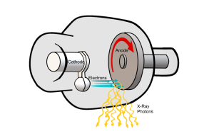
X-rays are a form of electromagnetic radiation produced when high-energy electrons interact with matter. In diagnostic and therapeutic settings, X-rays are generated within an X-ray tube when electrons accelerated by a high voltage strike a target material (usually tungsten). Understanding this process is essential for FRCR Part 1 Physics.
Two distinct mechanisms produce X-rays: bremsstrahlung radiation and characteristic radiation. Together, they create the X-ray spectrum used in medical imaging and radiotherapy.
The X-Ray Tube: Basic Components
An X-ray tube consists of:
- Cathode: A heated tungsten filament that releases electrons through thermionic emission
- Anode: A tungsten target that electrons strike to produce X-rays (rotating in diagnostic tubes to dissipate heat)
- Vacuum: The tube is evacuated so electrons don't collide with air molecules
- High voltage supply: Creates the electric field that accelerates electrons from cathode to anode (measured in kVp)
Why tungsten? Tungsten is used because of its high atomic number (Z=74), which increases X-ray production efficiency, and its high melting point (3,422°C), allowing it to withstand the heat generated. Only about 1% of electron energy is converted to X-rays—the remaining 99% becomes heat.
Bremsstrahlung Radiation
Bremsstrahlung (German for "braking radiation") accounts for approximately 80% of X-rays produced in a diagnostic tube. It occurs through the following mechanism:
- A high-energy electron from the cathode passes close to a tungsten nucleus in the anode
- The electron is deflected and decelerated by the attractive force of the positively charged nucleus
- The kinetic energy lost during this deceleration is emitted as an X-ray photon
- The electron continues on a deflected path with reduced energy
Key Properties of Bremsstrahlung
- Continuous spectrum: X-ray photons are produced with a range of energies from zero up to the maximum energy of the incident electrons
- Maximum photon energy: Equals the peak tube voltage (kVp). For example, a 100 kVp tube produces X-rays with maximum energy of 100 keV
- Energy distribution: Low-energy photons are more numerous; the number of photons decreases with increasing energy
- Dependence on Z: The probability of bremsstrahlung production varies with Z² of the target material
Clinical note: At low electron energies (diagnostic radiology), bremsstrahlung X-rays are emitted in all directions. At high electron energies (linear accelerators in radiotherapy), X-rays are emitted preferentially in the forward direction.
Characteristic Radiation
Characteristic radiation accounts for approximately 20% of useful X-rays in a diagnostic tube (at typical kVp settings). The mechanism is:
- An incident electron knocks an inner-shell electron (usually K-shell or L-shell) out of a tungsten atom
- This creates a vacancy in the inner shell
- An electron from a higher energy shell drops down to fill the vacancy
- The energy difference between shells is released as an X-ray photon
Key Properties of Characteristic Radiation
- Discrete energies: X-ray photons are produced at specific energies corresponding to the binding energy differences between electron shells
- Material dependent: The characteristic energies depend on the atomic structure of the target material, not the tube voltage (hence "characteristic")
- Threshold energy: Incident electrons must have at least the binding energy of the inner shell to eject an electron
Tungsten Characteristic X-Ray Energies
For tungsten (K-shell binding energy ≈ 69.5 keV):
- Kα radiation: ~59.3 keV (L→K transition)
- Kβ radiation: ~67.6 keV (M→K transition)
Characteristic X-rays from tungsten only appear in the spectrum when the tube voltage exceeds ~70 kVp (above the K-shell binding energy).
The X-Ray Spectrum
The complete X-ray spectrum is a combination of:
- The continuous bremsstrahlung spectrum (shaped like a ramp, decreasing from low to high energies)
- Characteristic peaks superimposed at specific energies
Factors Affecting the X-Ray Spectrum
Tube Voltage (kVp)
- Increasing kVp increases the maximum photon energy
- Increases the quantity of X-rays produced
- Increases the average energy (beam quality)
- X-ray quantity is proportional to kVp²
Tube Current (mA)
- Increasing mA increases the number of electrons hitting the target
- Increases X-ray quantity proportionally
- Does not change the spectrum shape or maximum energy
Target Material (Atomic Number)
- Higher Z materials produce more X-rays (efficiency ∝ Z)
- Changes the characteristic peak positions
Filtration
- Filters (usually aluminium in diagnostic imaging, higher Z materials in therapy) preferentially absorb low-energy X-rays
- This "beam hardening" increases the average beam energy
- Reduces patient skin dose by removing photons that wouldn't penetrate to the detector
- Minimum total filtration of 2.5 mm aluminium equivalent is required for diagnostic tubes >110 kVp
Comparison: Diagnostic vs. Therapeutic X-Ray Production
| Feature | Diagnostic | Therapeutic (Linac) |
|---|---|---|
| Electron energy | 25-150 keV | 4-25 MeV |
| Target type | Thick target at 90° | Thin transmission target |
| X-ray direction | All directions | Forward directed |
| Characteristic radiation | Significant contribution | Negligible (high energies) |
Key Points for FRCR Part 1
- Bremsstrahlung produces a continuous spectrum; characteristic radiation produces discrete peaks
- Only ~1% of electron energy is converted to X-rays; 99% becomes heat
- Maximum photon energy equals the tube voltage (kVp)
- X-ray quantity ∝ kVp² × mA × Z
- Filtration removes low-energy photons, increasing average beam energy
- Characteristic radiation from tungsten only appears above ~70 kVp
Practice Physics with PassOncology
Our Physics module covers X-ray production, radiation interactions, dosimetry, treatment planning, and all other key topics for FRCR Part 1. Build confidence with our comprehensive question bank.
Enjoyed this article?
Share it with a colleague preparing for their FRCR.